Cell- and tissue-specific detection of jasmonates
A gap in our knowledge on jasmonate-induced processes is the cellular localization of jasmonates. Most functions of jasmonates within distinct cells or tissues have been identified through indirect analyses using localization of JA biosynthetic enzymes and JA-induced proteins. To visualize JA directly in cross sections of plant material, we developed a novel antibody-based approach. The antibodies raised in rabbit against jasmonic acid (JA) are specific for bioactive jasmonates, such as JA, its methyl ester and isoleucine conjugate. In combination with newly established fixation and embedding methods, JA was detected by immunolabeling within all cells near the wound site of a mechanically wounded tomato leaf (Mielke et al., 2011). In a second approach, we aim to develop a non-invasive method for the visualization of JA. Using the promoters of the highly JA responsive AOC2 and AOC3 genes of Arabidopsis thaliana, JA-specific cis-elements will be selected and used for the construction of a synthetic promoter. Promoter activity will be monitored through GFP fluorescence in transient (Arabidopsis protoplasts, leaves of Nicotiana benthamiana) and stable (plants) transformation systems. In collaboration with Dr. S. Marillonnet we aim to use TALE-based systems for monitoring cell-specific occurrence of jasmonates.
Mielke, K., Forner, S., Kramell, R., Conrad, U., and Hause, B. (2011). Cell-specific visualization of jasmonates in wounded tomato and Arabidopsis leaves using jasmonate-specific antibodies. New Phytol. 190, 1069-1080.
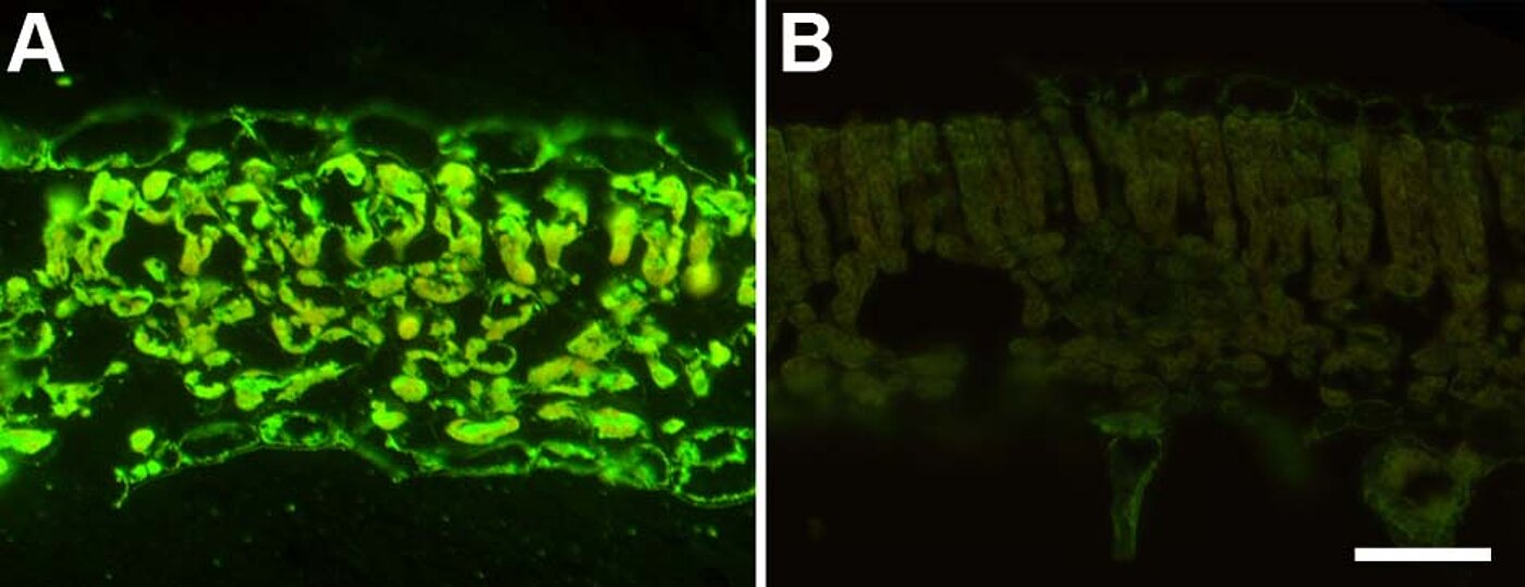
Fig. 1: Immunological detection of jasmonic acid in tomato leaves after wounding. Leaves of tomato wild type (A) or of the mutant acx1, which is JA deficient (B), were wounded, fixed and processed for immunolabeling using a JA-specific antibody. The green fluorescence visualizes the occurrence of JA and is visible in all epidermal and mesophyll cells of the wild type leaf, but is absent from the mutant leaf. Bar represents 50 µm for both micrographs. From Mielke et al. (2011).
This page was last modified on 27 Jan 2025 27 Jan 2025 .