Imaging Unit
Nowadays, the elucidation of molecular and biochemical processes requires its combination with investigation of cells – their physiological properties, their structure, the organelles they contain, interactions with their environment, their life cycle, division and death. For centuries, progress in biological research has been connected to the development of tools and equipment that allow new insights into living matter. The invention of and improvements in optical systems were very important because exceeding the limits of the optical resolution of the human eye delivered new insights into tissues, cells and subcellular compartments on the one hand and cellular processes on the other.
The Imaging Unit of the IPB aims to support all research groups in their use of cell biological methods. Currently, at least 13 research groups of the institute are using this unit.
The unit provides:
- coordinated supervision and maintenance of equipment
- optimal training of coworkers
- maximal use of IPB investments
- updating of equipment according to state-of-the-art and to requirements of actual research
The working principle of this unit is:
- unit headed by one scientist (Prof. Dr. Bettina Hause) and supported by one technician (Hagen Stellmach)
- equipment is located decentralized, but its maintenance is carried out centralized
- for all cell biological methods advice, training and help is provided, extensive experiments, however, have to be done by the co-workers themselves
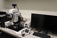
Microscopes
Epi-fluorescence microscopes
Confocal Laser Scanning Microscope
More devices
- Organelle-marker: Vectors and transgenic lines of Arabidopsis (Nelson et al., 2007)
- Wave-marker: Vectors and transgenic lines of Arabidopsis (Geldner et al., 2009)
- Golden-Gate-based Organelle-marker (Stellmach et al., 2022)
Methods
- Fixiation, embedding and sectioning of plant materials
- Laser-Micro-Dissection
- Immuno labelling
- in situ-hybridisation
- light microscopy including fluorescence microscopy
- confocal laser scanning microscopy
- Determination of protein interactions via FRET and BiFC (Split-YFP)
This page was last modified on 13 Mar 2025 13 Mar 2025 13 Mar 2025 13 Mar 2025 13 Mar 2025 13 Mar 2025 13 Mar 2025 13 Mar 2025 13 Mar 2025 13 Mar 2025 13 Mar 2025 13 Mar 2025 13 Mar 2025 13 Mar 2025 .


